

Structure of myosin-1c tail bound to calmodulin provides insights into calcium-mediated conformational coupling
2014.12.01Lu, Q., Li, J., Ye, F., & Zhang, M. (2015). Nature structural & molecular biology, 22(1), 81-88.
Class I myosins can sense cellular mechanical forces and function as tension-sensitive anchors or transporters. How mechanical load is transduced from the membrane-binding tail to the force-generating head in myosin-1 is unknown. Here we determined the crystal structure of the entire tail of mouse myosin-1c in complex with apocalmodulin, showing that myosin-1c adopts a stable monomer conformation suited for force transduction. The lever-arm helix and the C-terminal extended PH domain of the motor are coupled by a stable post-IQ domain bound to calmodulin in a highly unusual mode. Ca2+ binding to calmodulin induces major conformational changes in both IQ motifs and the post-IQ domain and increases flexibility of the myosin-1c tail. Our study provides a structural blueprint for the neck and tail domains of myosin-1 and expands the target binding modes of the master Ca2+-signal regulator calmodulin.
- Recommend
-
2025-10-22
IQSEC2/BRAG1 may modulate postsynaptic density assembly through Ca2+-induced phase separation.
-
2025-08-22
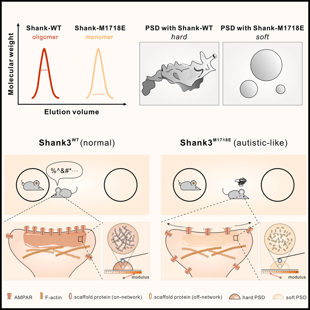
Shank3 oligomerization governs material properties of the postsynaptic density condensate and synaptic plasticity.
-
2025-08-21
Modulating synaptic glutamate receptors by targeting network nodes of the postsynaptic density condensate.
-
2025-08-19
Current practices in the study of biomolecular condensates: a community comment.
-
2025-06-10
Phase separation instead of binding strength determines target specificities of MAGUKs.